

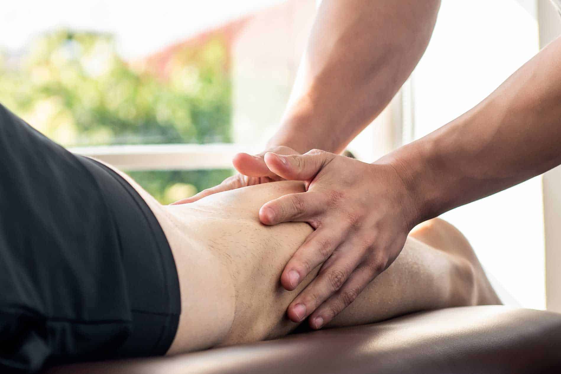
If you come to Blackberry Clinic with a painful musculoskeletal condition you might be told by your practitioner that you have ‘ fasciitis’ or that your ‘ fascia is tight’. What is fascia? Why does it get so painful? What can you do about painful fascia? Inga Nez, one of our osteopaths at Blackberry Clinic Milton Keynes has put together a very comprehensive article explaining what fascia is, what the structure of fascia is and why it can become painful. In addition, she covers treatment options that you might have if your fascia is causing an issue.
About fascia
To understand fascia and its importance in health, it’s necessary to comprehend its structure, morphology and function.
FASCIA
A preliminary definition of the fascial system was completed in September 2016.
The fascial system interpenetrates and surrounds all organs, muscles, bones and nerve fibers, endowing the body with a functional structure, and providing an environment that enables all body systems to operate in an integrated manner.
The fascial system consists of the three-dimensional continuum of soft, collagen-containing, loose and dense fibrous connective tissues that permeate the body. It incorporates elements such as adipose tissue, adventitiae and neurovascular sheaths, aponeuroses, deep and superficial fasciae, epineurium, joint capsules, ligaments, membranes, meninges, myofascial expansions, periostea, retinacula, septa, tendons, visceral fasciae, and all the intramuscular and intermuscular connective tissues including endo-/peri- /epimysium.(3)
STRUCTURE
In an operational simplicity, it is a sheet of connective tissue that surrounds, attaches, interlinks and separates internal organs and skeletal muscles. Houses blood and lymph vessels, nerves, and molecules able to modulate physiological processes.
It has 3 layers:
1. Superficial fascia, under the skin, surrounding the whole body like the second inner skin.
2. Deep fascia
a) epimysium, aponeurosis: surrounding the muscles, its continuation are tendons and ligaments.
b) intermuscular: surrounding the muscle fibers
3. Parietal and visceral fascia surrounds the viscera (stomach, lung, liver, heart, nerves, etc.) of the body.
In between these layers there is fluid film, mainly of hyaluronic acid and hondroitin sulphate, which ensures layers ability of smooth glide during movement.
The use of new research technologies, such as endoscopic micro-videography, reveals fascia as a ‘chaotic fibrillar network,’ with a dynamically changeable 3-dimentional architectural structure. (3)
Fascial tissues in those leyers differ in terms of their density and directional alignment of collagen fibers. E.g. superficial fascia is characterized by a loose density and mostly multidirectional or irregular fibre alignment. Whereas in the denser tendons or ligaments the fibres are mostly unidirectional. The intramuscular fasciae ( septi, perimysium and endomysium) may express varying degrees of directionality and density. The same is true visceral fasciae. there are substantial overlaps areas in which a clear tissue category will be difficult or arbitrary.(2)
MORPHOLOGY of fascial tissue
On a histological level, the fascial system is constituted from several, rather than just one, types of connective tissue – e.g., areolar, dense regular/irregular, adipose. (3)
1.Fibroblast (make and secrete all fibers of areoal connective tissue)
2.Collagen fibers (strongest and most abundant fibers)
3.Ellastic fibers ( rubber like fibers which allow tissue to return to its original shape)
4.Reticular fibers (connect vessels and nerves, and fascial layers perpendicularly)
5.Ground substance ( extracellular matrix that holds interstitial fluid via sugar-protein molecules (hyaluronic acid and hondroitin sulphate), acts like a sponge. When there is increased inflammatory response it becomes more viscous, sticky)
PHYSIOLOGY of fascial tissue
Fascia is a living tissue.There is a constant molecular crosstalk between extracellular matrix (ECM) molecules and cells, and their components.
Small functional and structural alterations in the ECM result in complex cellular adaptation processes and, vice versa, changes in cell function and structure lead to ECM adaptation.(4)
All factors influencing cell or ECM behaviour like: exercise, injury, blood supply, hydration, nutrition can result in changes in the structure and homeostasis of tissues and organs.
The ECM also works as a molecular store, catching and releasing biologically active molecules to regulate tissue and organ function, growth and regeneration.
Various factors can have impact on fascia physiological state, for example: mechanical stress, hormones, age.
Mechanical stress can induce the release and activation of ECM-stored molecules, which in turn can modulate vascular growth and energy expenditure. Physical activity and exercise ( acute and chronic loading) stimulate collagen remodelling in tendons.
In elderly, postmenopausal women: oestrogen replacement impairs collagen synthesis in response to exercise, but has a stimulating effect on collagen synthesis at rest. Oral contraceptives have an overall depressing effect on collagen synthesis.
Age-related alterations in fascial tissues include: densification (alterations of loose connective tissue) and fibrosis (alterations of collagen fibrous bundles). Ageing is characterised by chronic, low-grade inflammation—the so-called inflammaging.
FUNCTION of fascial tissue
Fascia plays very important functions in the body, including but not limited to architectural/structural, neurological biomechanical force transmission, morphogenesis, and cellular signal transmission .
Mechanical
1. Scaffolding – supports structure of the body, its organs and systems. Sepparates, protects, connects.
2. Movement- connects musccles and optimises transfer of muscle forces and shock absorbtion.
Tendons are main components of force transmission but intermuscular and extramuscular fascial tissues also provide a pathway for force transmission, and co-creates so called kinetic chains.
Kinetic chains. Because of multidirectional network of myofascial continuity, altered local forces (eg. muscular contraction) also affect the mechanics of adjacent tissues, both near and far. For example:
· the knee-joint capsule is influenced by directly inserting tendons and by more distant structures such as the gluteus maximus or the tensor fasciae latae, latissimys dorsi, and their connecting fasciae.
· stretching of the lower limb increases the range of motion of the cervical spine
· patients with sacroiliac pain display hyperactivity of the gluteus maximus and the contralateral latissimus dorsi. (2)
3. Fluid mobility
Fascia supports smooth mobility of different layers of muscles, bones, viscera, nerves and vessels.
Nutrition, Healing, Information
Fascia is housing nerve fibers, blood and lymph vessels.
On the fascial healthy structure and function depends proper vascular function (nutrition, cleansing, healing, hormons) of the neigbouring tissue, and transmission of neural information (proprioception, tension, pressure, injury) between the peripheral and central nervous system.
Some molecules embedded in fascia can influence/modulate such processes as growth, healing remodelling and inflammation. (Tenascin and osteopontin are examples of ECM molecules important for the regulation of the local immune response).
In addition, ECM plays an important role as a barrier to transmigration of immune cells in and out of the tissue.
INJURY of fascial tissue
Injuries to the fascial system can cause a significant loss of quality of physical performance.And can have a potential role in the development and perpetuation of musculoskeletal disorders, including lower back pain.(1)
Excessive or prolonged loading (exercise, work, and other activities) or direct trauma to fascial tissues initiates micro and macro changes necessary for tissue repair. Repairing response can become equally excessive and as such lead to:
Fibrotic changes -collagen deposition – tissue becomes dense and less elastic, in other words – congested, so called knots or trigger points are being formed.
Compression – fibrosis around the tendon, nerve and myofascial tissues can tether structures to each other or induce chronic compression.
Sensitisation of tissue – repetitive stress, injury can cause excessive release of substance P (neuropeptide) responsible for sending nociceptive signal to Central Nervous System.
Secondary tissue damage-Inflammatory cytokines can ‘spill over’ into the bloodstream, leading to widespread secondary tissue damage and central nociceptor windup.
Changes in muscle fibre composition, adiposity and fibrosis in response to injury to related structures (eg, injury to an intervertebral disc) even in the absence of muscle trauma.
After an injury to an intervertebral disc, deep back muscles undergo rapid atrophy,most likely mediated by neural changes such as reflex inhibition.
This is followed by changes in muscle fibre composition (slow-to-fast muscle fibre transition),
fibrosis and fatty infiltration associated with increased production of proinflammatory cytokines (eg, TNF).
These effects may also contribute to pathological changes that modify tissue function and mechanics, leading to compromised performance. The effects may become systemic, and thus not limited to the injured/loaded tissues.
TREATMENT of fascial tissue
A major goal of clinicians is to return patients to activity, training and competition after injury.
Exercise, physical modalities and pharmacological interventions can reduce the inflammatory processes associated with fascial tissue injury and fibrosis.
For example:
Anti-inflammatory drugs. Early treatment with them can prevent pain behaviours induced by TNF signalling and reduce excessive collagen production. However, overuse of ati-inflammatory drugs-may impair regeneration and diminish tissue adaptation.
Stretching of fascial tissues can promote resolution of inflammation .
Exercise. Resistance exercise is necessary to reverse fatty changes (and perhaps fibrosis) in chronic conditions, whereas gentle muscle activation is sufficient to reverse early muscle atrophy. Whole body exercise can prevent inflammatory changes in back muscles that follow intervertebral disc injuries.
Injections of platelet-rich plasma seem to be successful in some cases of tendinopathy, although efficacy remains inconclusive.
Manual therapy (osteopathy, chiropractic, massage, cupping, foam rolling, shockwave therapy) can improve short term flexibility and recovery from muscle soreness , decrease latent trigger point sensitivity, enhance fascial layer sliding, modify corticospinal excitability, and prevent overuse-induced fibrosis.
References:
1) Zügel, M., Maganaris, C. N., Wilke, J., Jurkat-Rott, K., Klingler, W., Wearing, S. C., Findley, T., Barbe, M. F., Steinacker, J. M., Vleeming, A., Bloch, W., Schleip, R., & Hodges, P. W. (2018). Fascial tissue research in sports medicine: from molecules to tissue adaptation, injury and diagnostics: consensus statement. British journal of sports medicine, 52(23), 1497. https://doi.org/10.1136/bjsports-2018-099308
2) Schleip, R., & Müller, D. G. (2013). Training principles for fascial connective tissues: scientific foundation and suggested practical applications. Journal of bodywork and movement therapies, 17(1), 103–115. https://doi.org/10.1016/j.jbmt.2012.06.007
3) Adstrum, S., Hedley, G., Schleip, R., Stecco, C., & Yucesoy, C. A. (2017). Defining the fascial system. Journal of bodywork and movement therapies, 21(1), 173–177. https://doi.org/10.1016/j.jbmt.2016.11.003
4) Chen, B., Ji, B., & Gao, H. (2015). Modeling Active Mechanosensing in Cell-Matrix Interactions. Annual review of biophysics, 44, 1–32. https://doi.org/10.1146/annurev-biophys-051013-023102
5) Suhr, F., Brixius, K., & Bloch, W. (2009). Angiogenic and vascular modulation by extracellular matrix cleavage products. Current pharmaceutical design, 15(4), 389–410. https://doi.org/10.2174/138161209787315756
6) Carla Stecco’s Functional Atlas of the Human Fascial System (2015
Written by Inga Nez
Osteopath, Blackberry Clinic Milton Keynes